Eye Case Studies — real clinical images, annotated
A growing library of anonymised patient cases from our clinic — slit-lamp, fundus, OCT, FFA and topography imaging with the same interpretation and teaching notes we share with our post-graduate students. Educational only; not a substitute for a formal consultation.
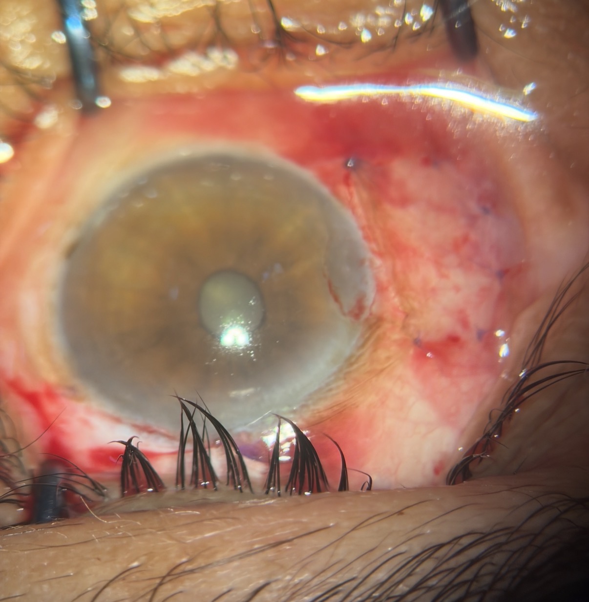
Pterygium (नाखूना) treatment using Limbal Conjunctival Autograft in Varanasi
LCAT (Limbal Conjunctival Autograft) is the best technique of pterygium surgery with almost nil recurrence rates. Shantanu Netralya offers this specialised treatment in a seamless manner with no injection no pain technique.
Wet AMD on OCT — subretinal fluid and pigment epithelial detachment
Classic OCT signs of neovascular (wet) AMD before the first anti-VEGF injection.
Mature white cataract — slit-lamp view before MICS phacoemulsification
Dense white cataract with no red reflex — a surgical planning case for capsular staining and iris hooks.
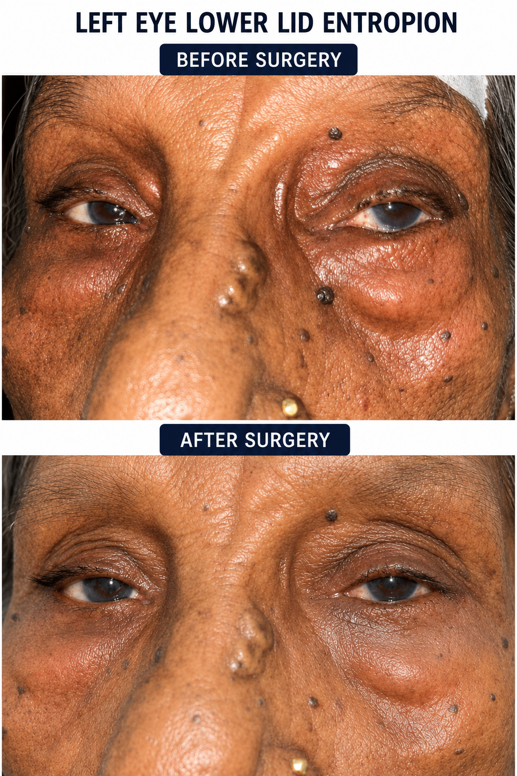
Entropion surgery
Patient complained of chronic irritation left eye since a long time
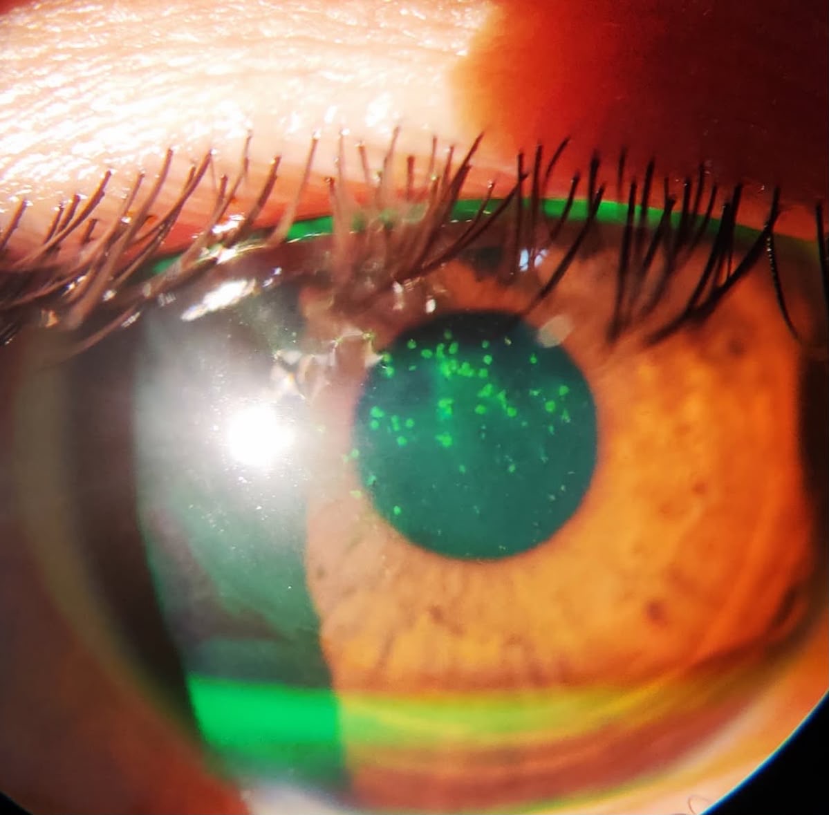
Superficial punctate keratitis post conjunctivitis
Superficial punctate keratitis post conjunctivitis
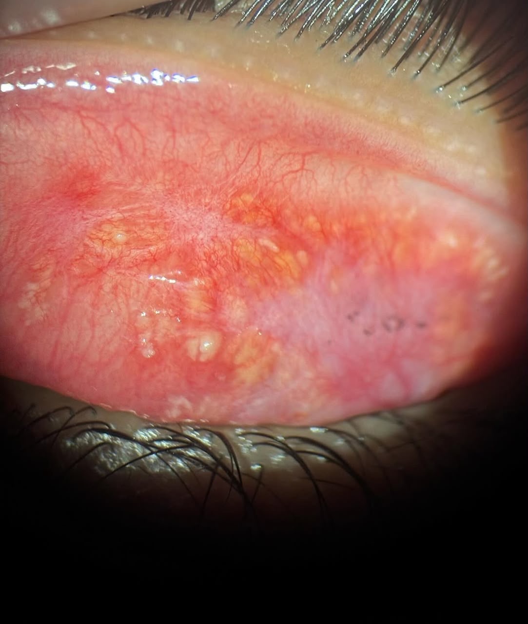
Concretions in eye
A simple lid eversion revealed why this patient had foreign body sensation for so long
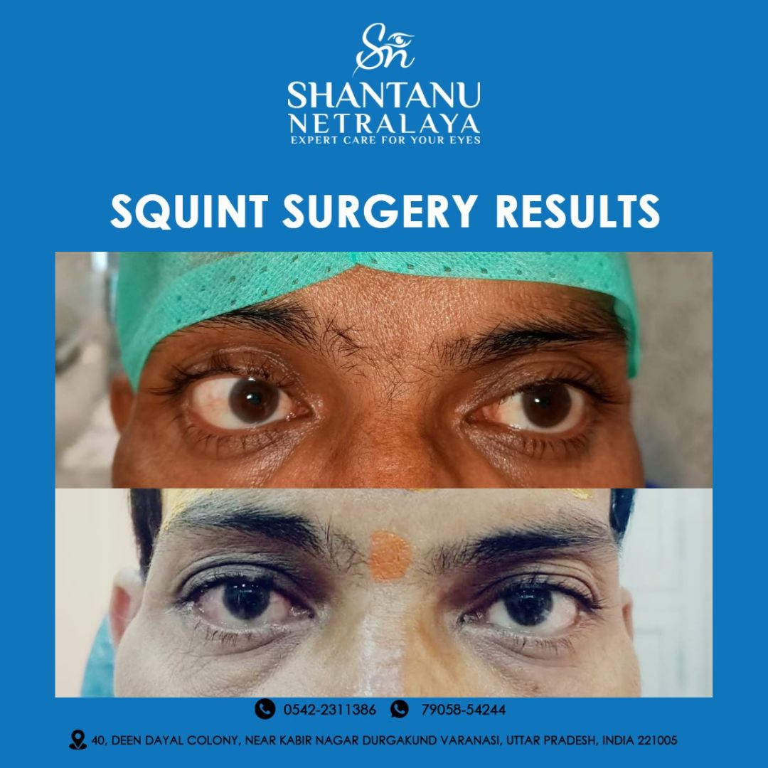
Paralytic squint treatment
Long standing 6th nerve palsy treated surgically
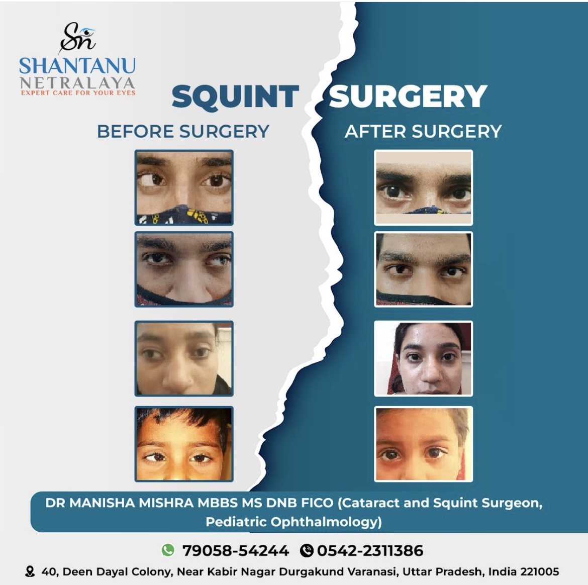
Life transforming squint surgery
Before and after images of some of our satisfied squint patients
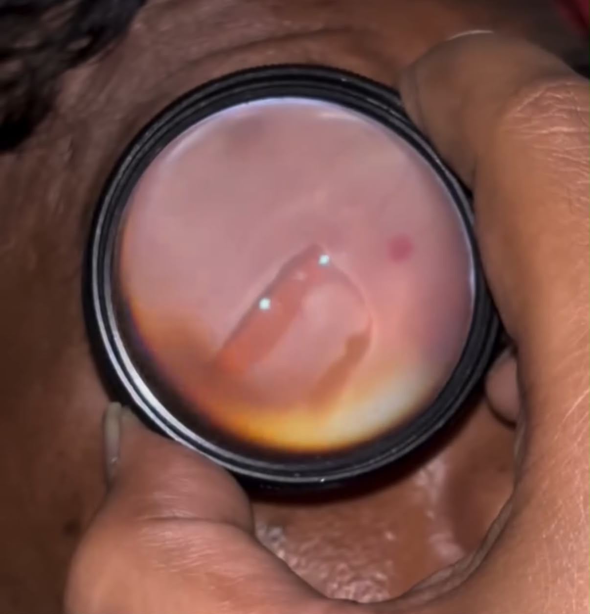
Catastrophic vision loss prevented
A large horse shoe tear in the retina
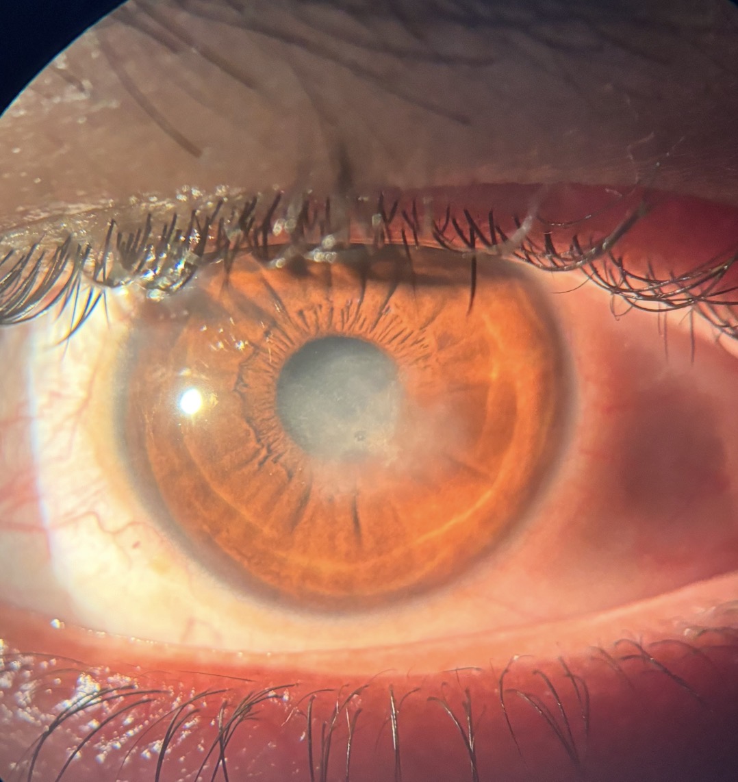
CORNEAL ENDOTHELITIS
Viral Infection of eye specifically the back part of the eye which causes corneal swelling and haziness in the cornea leading to poor vision, pain, photophobia
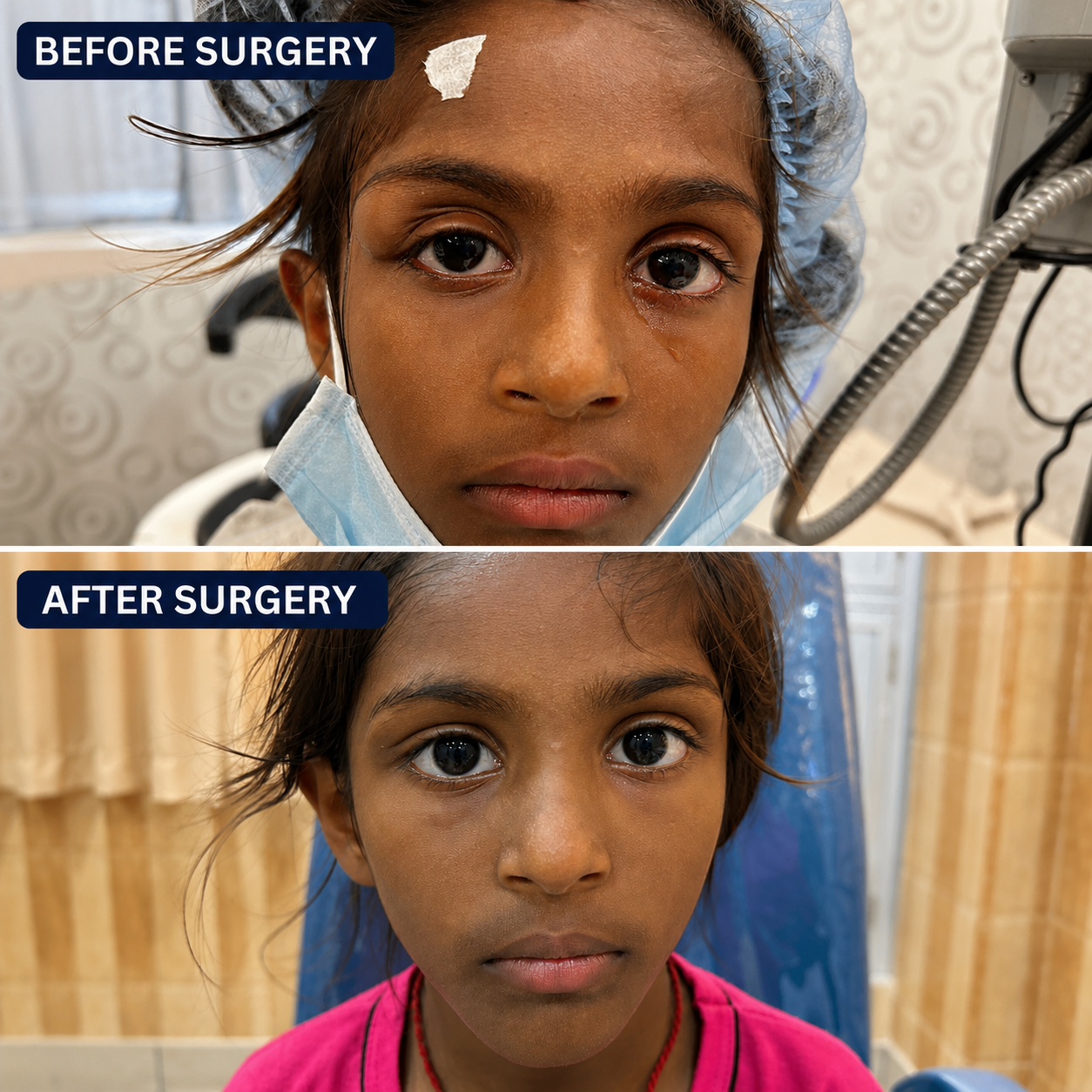
Orbital dermoid
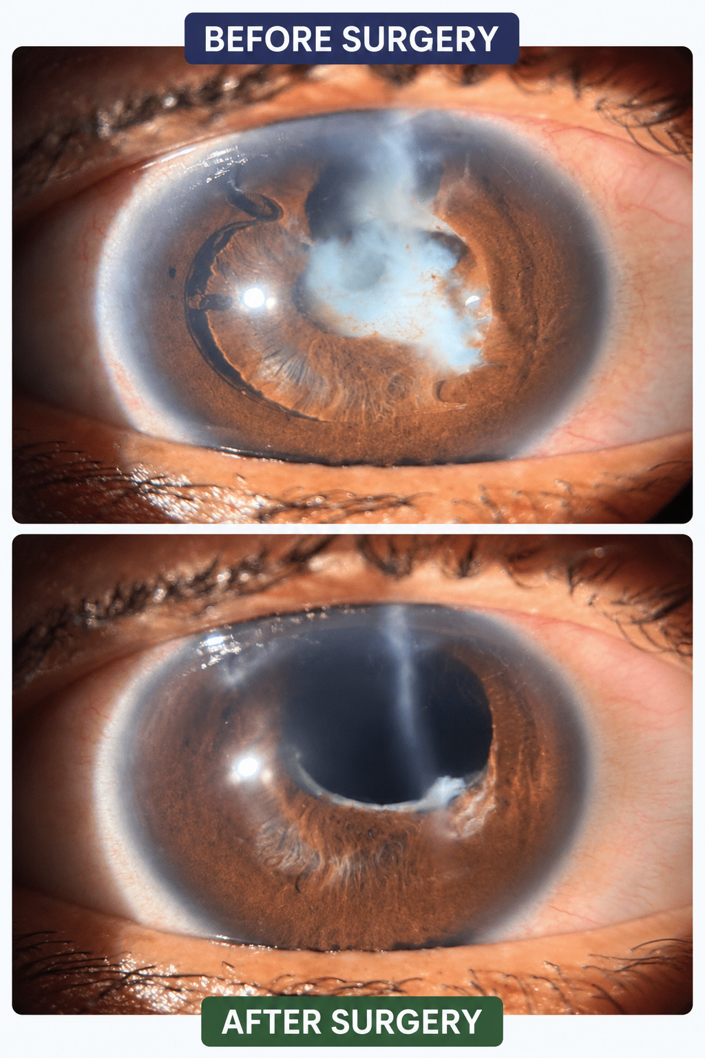
Complex re surgery
This patient was operated elsewhere in childhood for a post traumatic cataract. Over a span of several years the vision had worsened gradually. On examination we found complete opacification behind the lens. We accepted the challenge and took up the patient for surgery. Removed the lens and opacification and did an en extremely complex procedure of scleral fixated lens here at Shantanu Netralaya
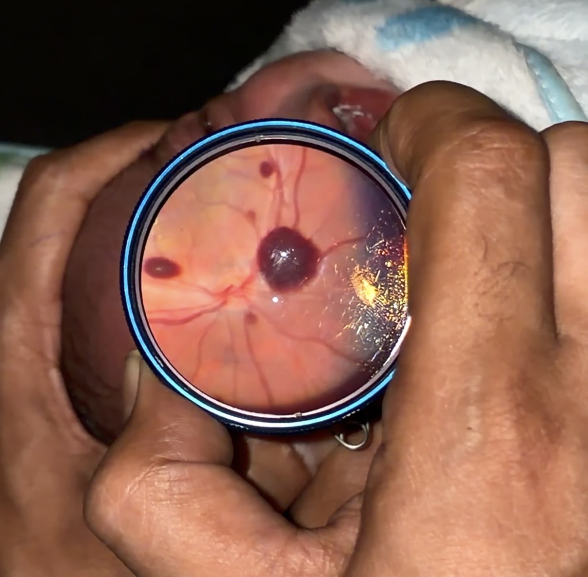
Birth Trauma-Related Retinal Hemorrhages
Birth trauma-related retinal hemorrhages are small areas of bleeding in the retina that can occur during a difficult or even normal vaginal delivery due to pressure changes during birth. They are relatively common in newborns, are not the same as Retinopathy of Prematurity (ROP), and in most cases resolve spontaneously within a few weeks without affecting vision.
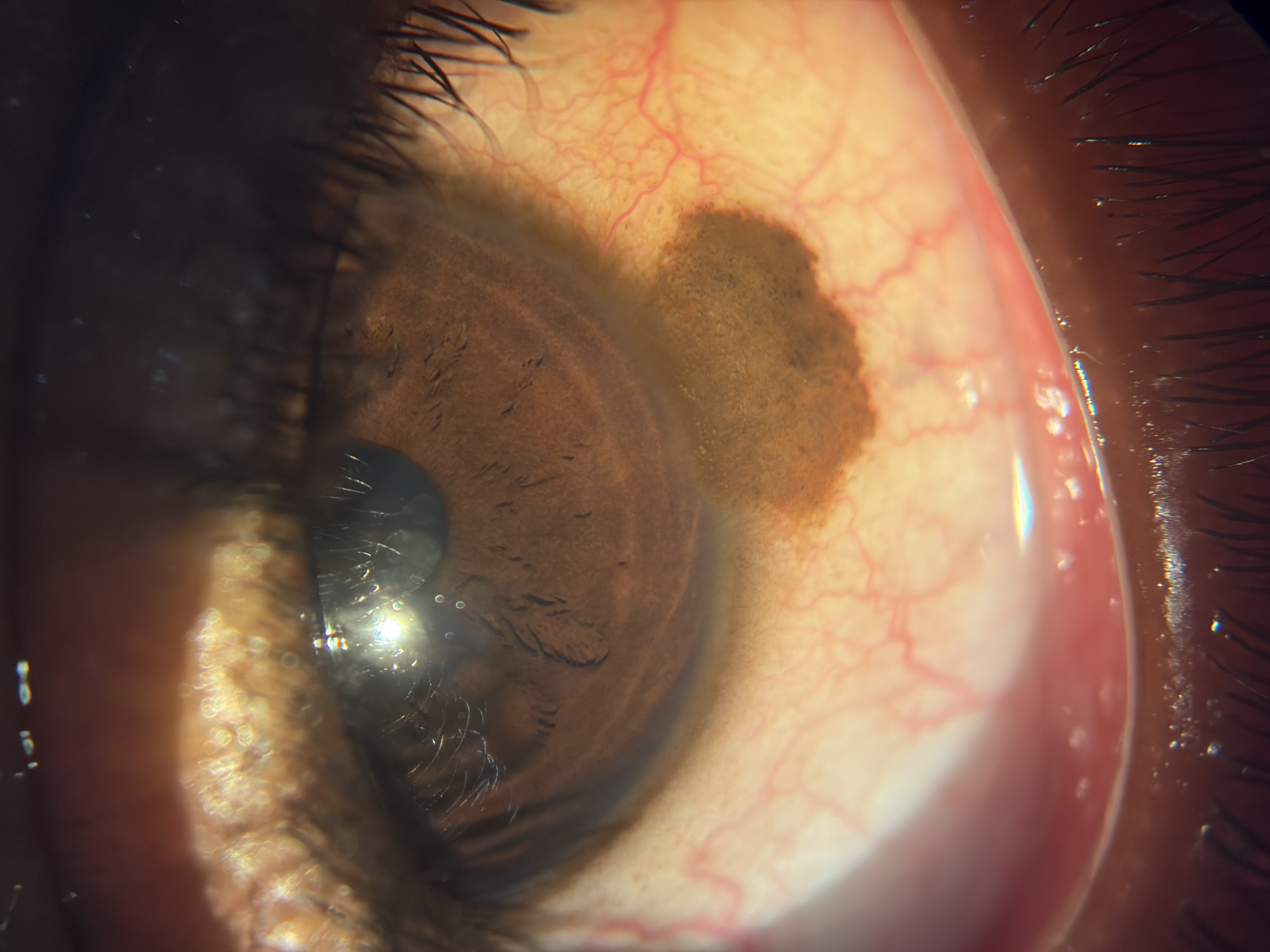
Conjunctival Naevus
A conjunctival nevus is a common, benign (non-cancerous) pigmented lesion of the conjunctiva, often referred to as a “mole on the eye.”
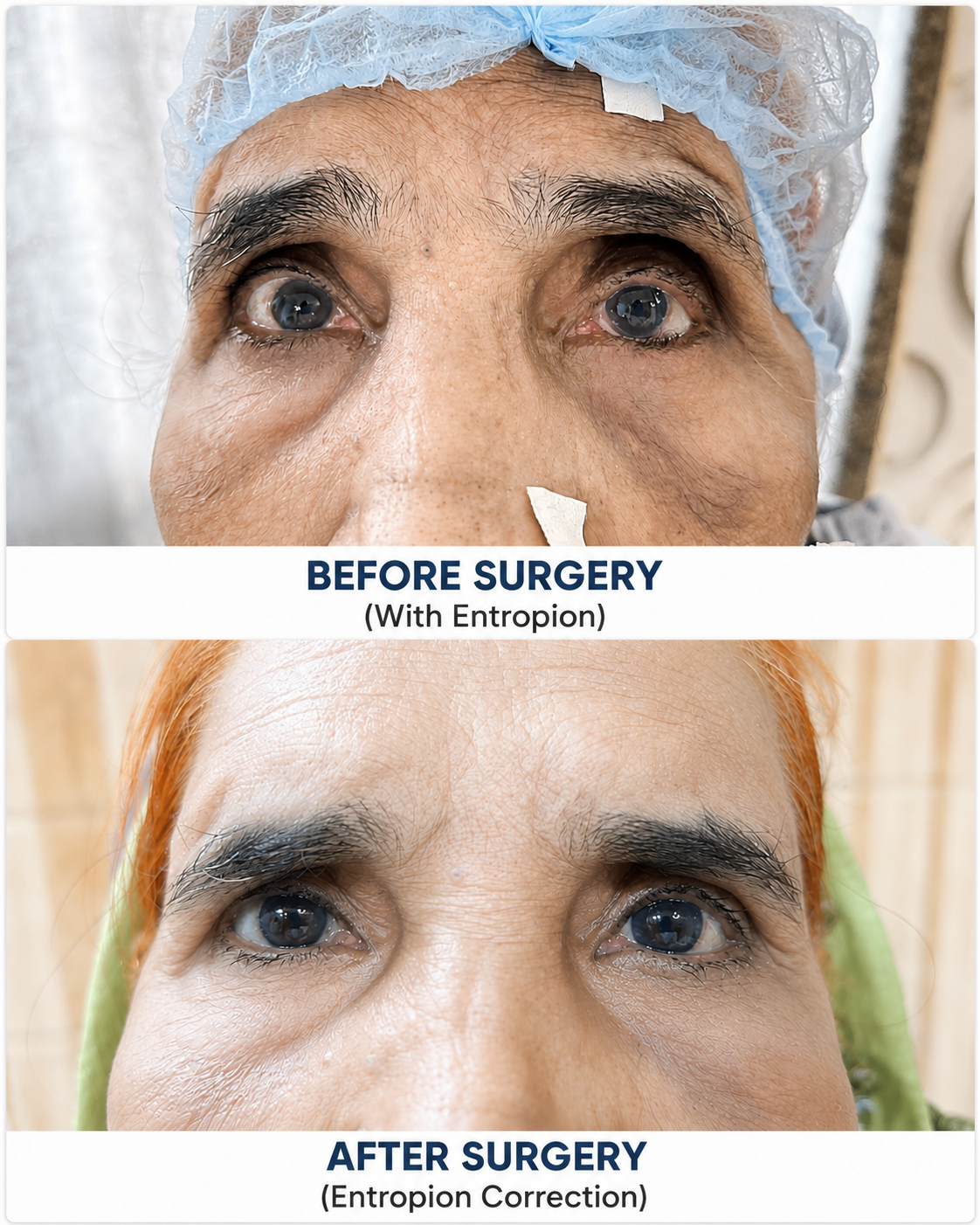
Entropion correction at Shantanu Netralaya
Left eye lower lid entropion correction with surgery
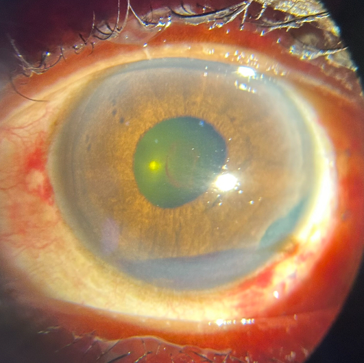
Ocular trauma
Patient presented with ocular trauma followed by pain and low vision
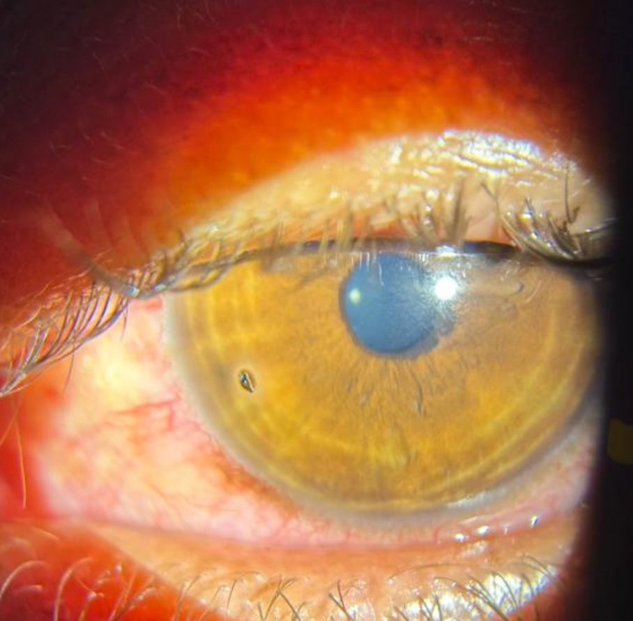
Superficial corneal foreign body
This picture shows a tiny splinter stuck on the cornea
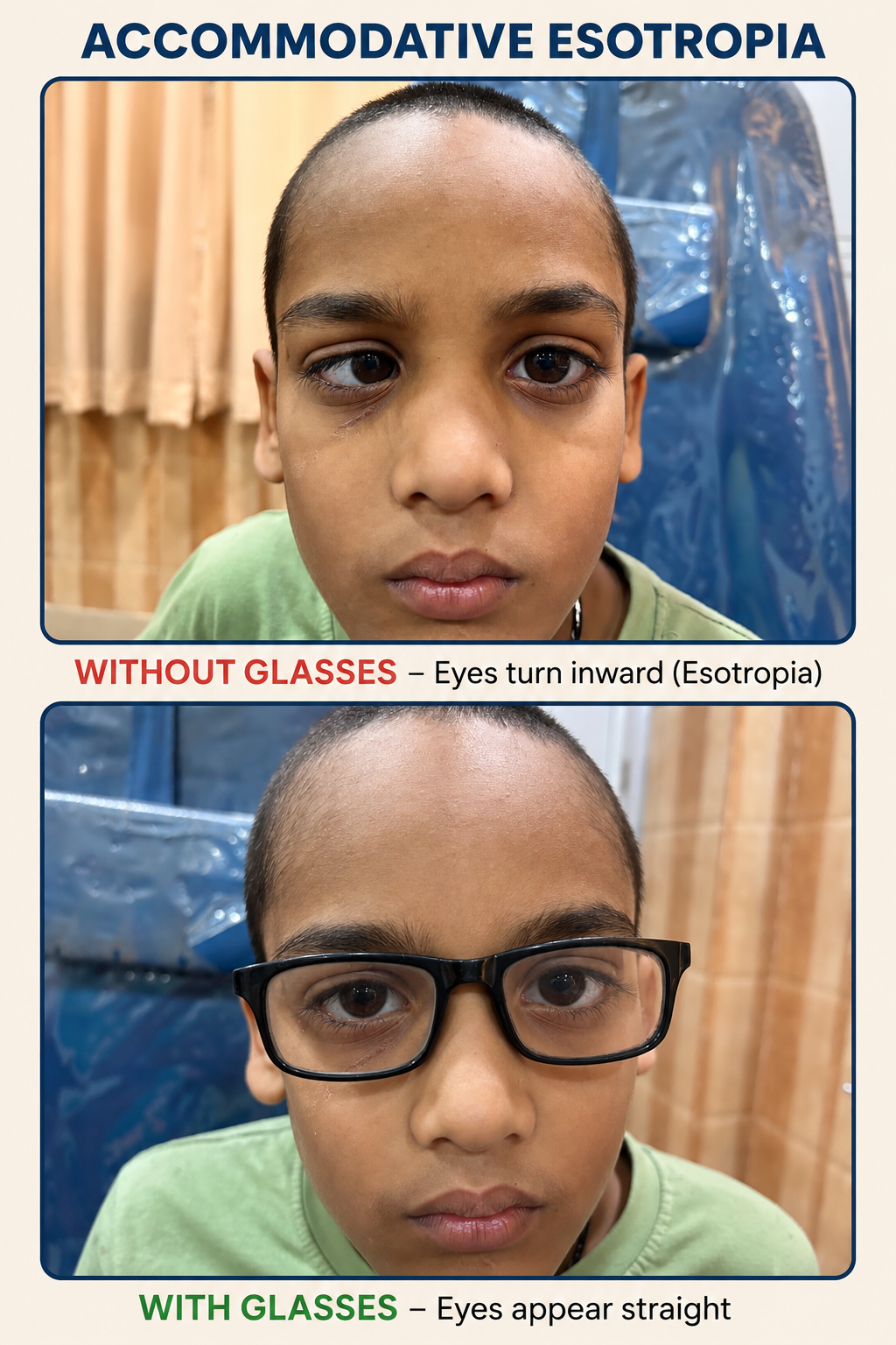
Accomodative esotropia
This picture shows a child whose squint is entirely corrected by glasses
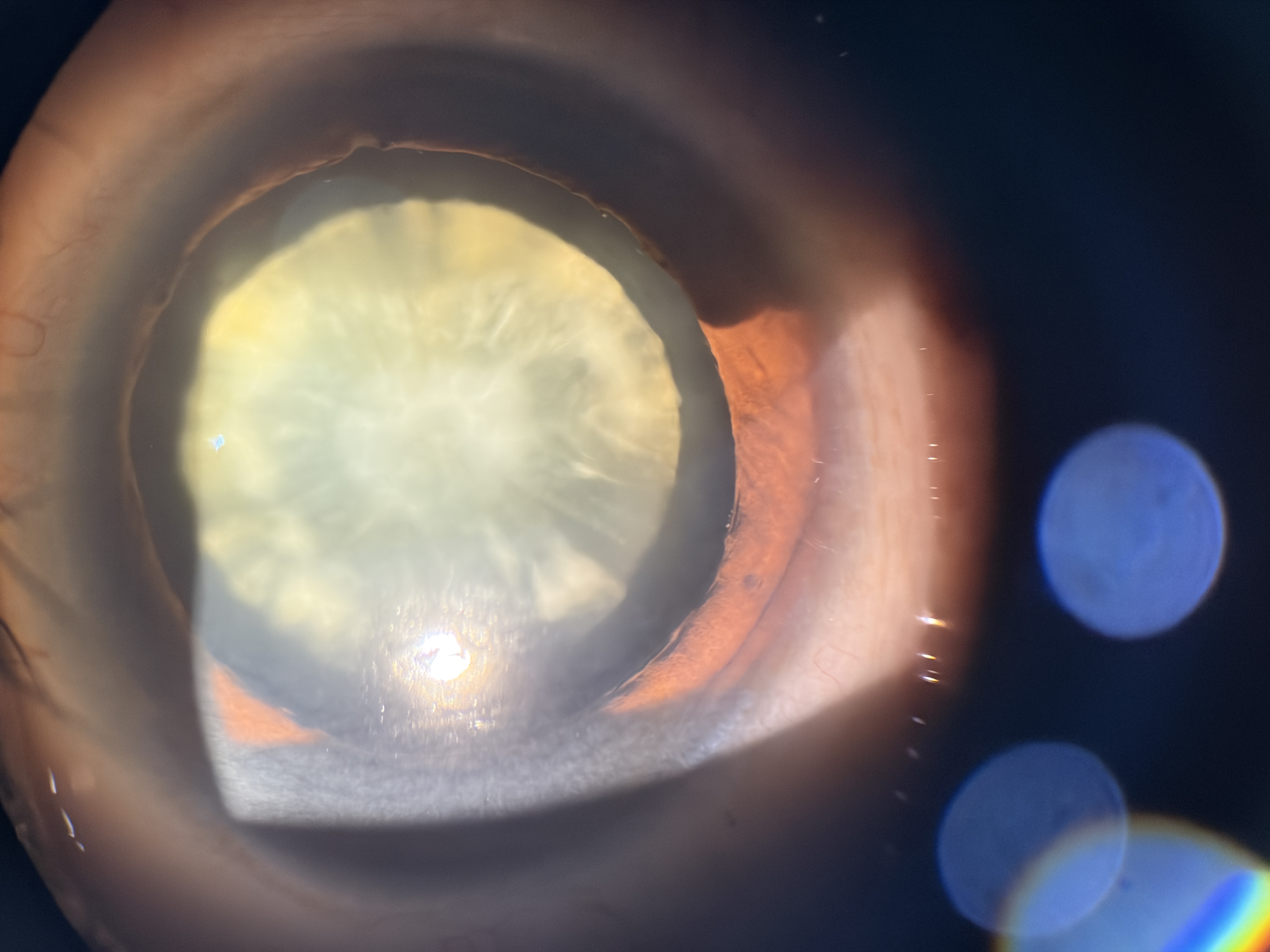
Zonular Cataract
Ignored case of zonular cataract now being planned for MICS Cataract Surgery in adulthood at 60 years of age
Severe non-proliferative diabetic retinopathy — colour fundus photograph
Multiple deep-blot haemorrhages, cotton-wool spots and venous beading in a 55-year-old type 2 diabetic.